التليف الرئوي احد اخطر مضاعفات الاصابة بكورونا COVID-19. للدكتور عروة محمد هاني الملي/المانيا
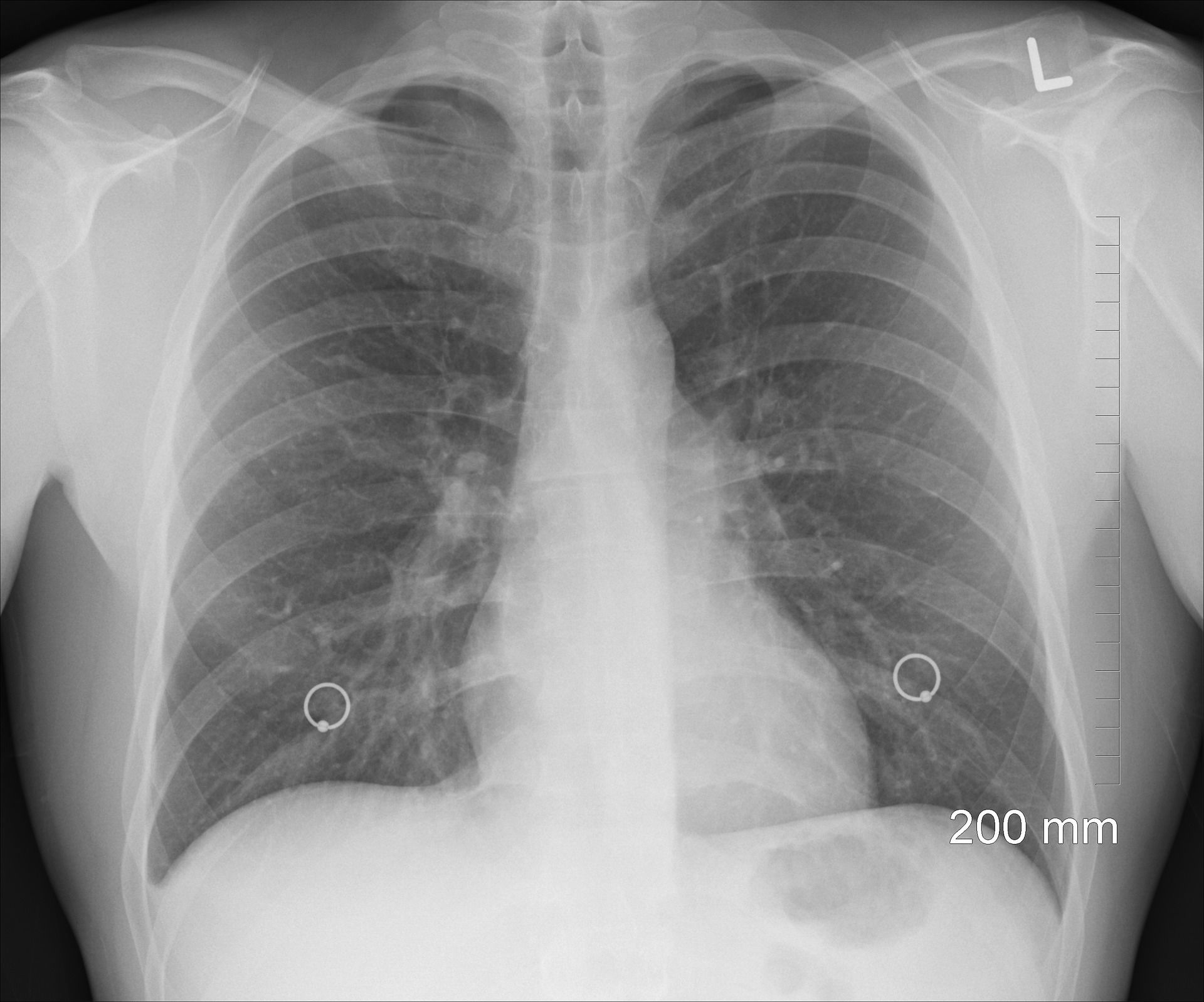
المضاعفات بعد اصابة كوفيد كثيرة و بحسب الدراسات المختلفة واشهرها ثلاث دراسات فإن 70 الى 80 % من المصابين يبقى لديهم عرض مزمن على الاقل بعد الشفاء. الدراسة الايطالية فصلت حتى النسب المئويه للاعراض المختلفة وهي موجودة ضمن المراجع للاطلاع. دراسة صينية نشرت الشهر الماضي اظهرت ان حوالى 8 % من المصابين يحدث لديهم تليف رئوي يجب التدخل بايجاد حل له قبل ان يحتاج المريض الى زراعة رئة يوما ما. ماهو التليف الرئوي من وجهة نظر الباثولوحيا هو حالة مزمنة يتشكل فيها الياف ضامة ونسيج ضام قاسي نسبيا في النسيج الخلالي الرئوي و جدار الاسناخ و الاوعية الدموية. طبعا بدل الشفاء الطبيعي الكامل يحدث نتيجة الدمار الكبير في نسيح الرئة نوع من الاصلاح بدل التجديد للنسيج المصاب وشفاؤه. الاصلاح يحدث عندما يكون الدمار اكبر من القدرة على التجديد او بمعنى عامي الخرق اكبر من الرقعة فيتم ردم النسيج المتهالك بدل تجديده. الردم يتم بتشكيل نسيج ضام في البداية على شكل خيوط في نسيج الرئة ولكن بحال كان الموات والدمار كبير جدا يتشكل ندب من النسيج الضام. هذه الندب ليس لها فعالية وظيفية بل على العكس تسبب انهاك في عمل الرئة بسبب قساوتها. ما اعراض التليف الرئوي . ضيق تنفس . في البداية يشعر المريض بالضيق عند الاجهاد والعمل ثم في حالة الراحه كمان ان البداية تظهر على شكل ضيق تنفس عند الزفير ثم عند الشهيق . سعال جاف الإرهاق فُقدان الوَزن وَجَع العضلات والمفاصل زيادة عرض ودوران أطراف أصابع الأيدي والأقدام (التعجُّر) الاعراض تختلف بشدة من شخص لآخر. حيث تظهر عند بعض المرضى اعراض شديدة بسرعة . يظهر آخرون أعراض متوسِّطة تزداد سوءًا وببطء خلال شهور إلى سنوات طبعا المرضى الذين احتاجوا عناية مشددة او تنفس اصطناعي هم اكثر عرضة للتليف الرئوي للاسف لايوجد علاج للتليف الرئوي واذا استمر من التقدم يصبح زراعة الرئة هي الحل الوحيد لانقاذ الانسان من الموت. د. عروة محمد هاني الملي الاكاديمية الالمانية العالمية للطب والابحاث Pulmonary fibrosis in critically ill patients with novel coronavirus pneumonia during the convalescent stage and a proposal for early intervention Hai Zou & Sheng-qing Li Acta Pharmacologica Sinica (2020)Cite this article Carfì A., Bernabei R., Landi F. For the Gemelli against COVID-19 post-acute care study group. Persistent symptoms in patients after acute COVID-19. J Am Med Assoc. 2020;324(6):603–605. [PMC free article] [PubMed] [Google Scholar] Istituto Superiore Sanità . 2020. Sorveglianza Integrata COVID-19 in Italia. [Google Scholar] Grasselli G., Zangrillo A., Zanella A. Baseline characteristics and outcomes of 1591 patients infected with SARS-CoV-2 admitted to ICUs of the Lombardy region, Italy. JAMA - J Am Med Assoc. 2020 doi: 10.1001/jama.2020.5394. [PMC free article] [PubMed] [CrossRef] [Google Scholar] Liu C., Ye L., Xia R. Chest CT and clinical follow-up of discharged patients with COVID-19 in Wenzhou City, Zhejiang, China. Ann Am Thorac Soc. 2020 doi: 10.1513/AnnalsATS.202004-324OC. [PMC free article] [PubMed] [CrossRef] [Google Scholar] Zhao Y.M., Shang Y.M., Song W.B. Follow-up study of the pulmonary function and related physiological characteristics of COVID-19 survivors three months after recovery. EClinicalMedicine. 2020;25:100463. doi: 10.1016/j.eclinm.2020.100463. [PMC free article] [PubMed] [CrossRef] [Google Scholar] . Huang C, Wang Y, Li X, Ren L, Zhao J, Hu Y, et al. Clinical features of patients infected with 2019 novel coronavirus in Wuhan, China. Lancet. 2020;395:497–506. Zou H, Li H, Zhang Y, Xia J, Zhang P, Xiong W, et al. Huashan model for the treatment of patients with severe novel coronavirus pneumonia and its clinical significance. Her Med. 2020;3:319–22. Google Scholar Diagnosis and Treatment Protocol for Novel Coronavirus Pneumonia (Trial Version 7) (Released by National Health Commission & National Administration of Traditional Chinese Medicine on March 3, 2020). Chin Med J (Engl). 2020;133:1087–95. Liu Q, Wang RS, Qu GQ, Wang YY, Liu P, Zhu YZ, et al. Gross examination report of a COVID-19 death autopsy. Fa Yi Xue Za Zhi. 2020;36:21–3 CAS،PubMed،Google Scholar He L, Wei M, Han Y, Zhang KJ, Yang Z, Wang SX. A study on association between SARS patients’ sequelae and HLA-A allele. Chin J Prev Med. 2009;10:825–8. Google Scholar Kong Q, Qin C. Comparative Study on pathogenesis of SARS pulmonary fibrosis. Chin J Comp Med. 2005;15:335–8.Google Scholar Xu Z, Shi L, Wang YJ, Zhang J, Huang L, Zhang C, et al. Pathological findings of COVID-19 associated with acute respiratory distress syndrome. Lancet Respir Med. 2020;8:420–2 Published: 13 November 2020 Pulmonary fibrosis in critically ill patients with novel coronavirus pneumonia during the convalescent stage and a proposal for early intervention Hai Zou & Sheng-qing Li Acta Pharmacologica Sinica (2020)Cite this article 1171 Accesses The coronavirus disease 2019 (COVID-19) outbreak poses a serious challenge to China and most countries around the world, thus the World Health Organization has declared the outbreak a public health emergency of international concern. On August 24, 2020, more than 23,584,000 cases of COVID-19 had been confirmed in more than 120 countries/regions worldwide, 812,519 patients had died, and 16,078,455 confirmed cases were still undergoing treatment. Most patients with COVID-19 have a good prognosis, but a small portion of patients become critically ill and even die (the cumulative mortality rate is ~3.7%). Critically ill patients account for ~15% of those with COVID-19, and most of them are the elderly, patients with underlying diseases or patients with obesity [1]. Critically ill patients often experience dyspnea and/or hypoxemia 1 week after onset of the illness, and in severe cases, the patient can rapidly develop acute respiratory distress syndrome (ARDS), septic shock, uncorrectable metabolic acidosis, bleeding and coagulation dysfunction, and multiple organ failure. According to our clinical treatment experience, we previously proposed the “Huashan Model”, which is based on supportive therapy for multiple organs supplemented with anti-inflammatory and anticoagulation therapies, for the treatment of patients with severe novel coronavirus pneumonia (NCP) [2]. This model significantly improved the survival rate of critically ill NCP patients by correcting the pathophysiological state, adjusting immune system function, and promoting clearance of the virus and affected cells through symptomatic and supportive treatment. Patients with severe NCP in the convalescent stage often experience serious complications, such as multiple organ failure. In particular, prevalent pulmonary parenchymal lesions, alveolar lumen exudates, and pulmonary interstitial fibrosis can lead to poor pulmonary function in patients and seriously affect their long-term quality of life. Therefore, alleviating/reversing the process of pulmonary interstitial fibrosis, improving the pulmonary function of patients with severe NCP, and improving the quality of life of patients should be the focus of treatment for patients with severe NCP in the convalescent stage. The pathological changes in the lungs of patients with NCP have the following characteristics. The lungs show varying degrees of consolidation, serous fluid and fibrinous exudates, and hyaline membrane formation is seen in the alveolar lumen. Exuded cells are mainly monocytes and macrophages, but multinucleated giant cells are also seen. The significant proliferation of type II alveolar epithelial cells is observed, and some cells are detached. Inclusion bodies are seen in type II alveolar epithelial cells and macrophages. Alveolar septal vessels exhibit congestion and edema, and mononuclear and lymphocytic infiltration and intravascular hyaline thrombus formation are observed. Focal hemorrhage and lung tissue necrosis are present, and hemorrhagic infarction may occur. Alveolar lumen exudates and pulmonary interstitial fibrosis are visible. Part of the epithelium of the bronchial mucosa in the lung detaches, and mucus and mucus plug formation are observed in the lumen [3]. A few alveoli are hyperinflated, and alveolar septa rupture and cyst formation are observed. Observation reports of the gross anatomy of deceased patients with NCP have revealed that the pleura is thickened and extensively adhered to lung tissues and that fibrous cords are visible in the lung section. The pathological features of NCP are very similar to those caused by severe acute respiratory syndrome (SARS) and Middle Eastrespiratory syndrome coronavirus (MERS), with pulmonary fibrosis and consolidation. The follow-up data for recovered SARS patients showed that 25.5%–62.0% of recovered SARS patients exhibited pulmonary interstitial fibrosis-like changes on 33–68 days after disease onset [4, 5]. Pulmonary fibrosis caused by SARS is not only one of the important clinical manifestations of the pathogenesis of the disease but also a more common sequela in SARS patients in the convalescent stage. Chest imaging has suggested that these patients have different degrees of fibrotic lesions. Currently, through the study of critically ill NCP patients successfully rescued by our team, the imaging findings of pulmonary fibrosis are as follows (Fig. 1): (1) reticular opacities accompanied by ground-glass opacities; (2) pulmonary consolidation combined with bronchiectasis; (3) reticular nodular opacities; and (4) fibrosis with other changes. Fig. 1: CT of CASE before and after treatment. figure1 An 80-year old woman 2 days after weaning ventilator with reticular nodule shadows and diffuse ground-glass opacities (GGO) in the two upper lobes (a); pulmonary consolidation with stretched bronchiectasis can be seen in the right lower lobe (b). A 70-year old man 2 days after weaning ventilator with consolidation, nodules and diffuse GGO in both lower lobes (c); after 1 week treatment with glucocorticoids (0.5 mg/kg every day), N-acetylcysteine and pirfenidone, most of lesions are absorbed with only reticular shadows left (d). Full size image Approximately 7%–8% of recovered patients have severe pulmonary fibrosis sequelae. It has been reported that lung injury in early-stage SARS patients involves a pathological change to alveolitis and induces the occurrence of fibrosis in later stages. Although most patients die of acute respiratory failure, as a sequela, the fibrosis that occurs at a later stage seriously affects pulmonary function and the quality of life of patients and leads to an impairment in the functions of other organs, which may lead to the death of SARS patients by secondary causes [6]. As a consequence, we suggest a close follow-up of recovered patients to find this sequela and an individualized treatment plan according to the degree of fibrosis. Both animal and human studies have shown that inflammatory monocytes-macrophages and neutrophils accumulate in the lungs after humans are infected by coronavirus. These cells are a major source of cytokines and chemokines associated with lethal human diseases caused by coronaviruses [7, 8]. Rapid viral replication and an exuberant proinflammatory cytokine/chemokine response lead to the apoptosis of lung epithelial cells and endothelial cells, which disrupts epithelial cell barriers of the pulmonary microvessels and alveoli, resulting in vascular leakage and alveolar edema and ultimately leading to hypoxia. The function and phenotype of pulmonary macrophages may vary depending on their polarization status, which is governed by the cytokine environment. The virus damages lung cells, causing the accumulation of macrophages that attempt to eliminate inflammation. However, rapid viral replication and the massive destruction of lung cells as well as an excessive inflammatory response instead induce M1-type polarization, thereby exacerbating pulmonary inflammation [9,10,11]. A study of the clinical characteristics of 99 patients with COVID-19 published in the journal Lancet found that the COVID-19 virus may act mainly on lymphocytes, especially T lymphocytes. Viral particles spread through the respiratory mucosa to infect other cells and induce a cytokine storm in vivo, and large amounts of plasma interleukin-2 (IL-2), IL-7, IL-10, granulocyte colony-stimulating factor (G-CSF), interferon (IFN)-γ, and monocyte chemotactic protein (MCP) are produced, causing the rapid progression of ARDS and septic shock in patients and eventually leading to multiple organ failure. Even surviving patients suffer from prolonged lung injury and fibrosis due to the excessive immune response, thus leading to respiratory dysfunction and a reduced quality of life [12]. At present, the main treatment is glucocorticoids. Due to excessive activation of the immune system and the rapid transformation in immunosuppression in critically ill patients, use of the hormones carries the risk of delayed viral clearance and secondary infection. Therefore, there is an urgent need to research and develop new intervention measures to inhibit excessive immune responses according to the disease course to protect alveolar function and reduce injuries to the lungs and systemic organs in patients with NCP. Currently, our treatment regimen for pulmonary fibrosis in patients with NCP is comprised of glucocorticoids (0.5–1 mg/kg every day), N-acetylcysteine, and pirfenidone in combination, which can accelerate the absorption of pulmonary shadows. Although there is no reliable evidence on the efficacy of corticosteroids in treating pulmonary fibrosis following SARS-CoV-2 infection, they are commonly used as an empirical therapy for postinflammatory pulmonary fibrosis on the basis of the patient’s clinical condition, High-Resolution Computed tomography manifestation and disease behavior. It could be suggested that pirfenidone, which shows effective against inflammation, fibrosis and oxidation [13], can attenuate lung injury because pirfenidone reduces LPS-induced acute lung injury and subsequent fibrosis [14]. Nintedanib can slow the progression of pulmonary fibrotic disease regardless of the pathological pattern by inhibiting the release of proinflammatory and profibrotic mediators, the migration and differentiation of fibrocytes and fibroblasts, and deposition of the extracellular matrix [15]. In light of these properties, pirfenidone and nintedanib might exert potential benefits in the prevention and treatment of pulmonary fibrosis following SARS-CoV-2 infection.